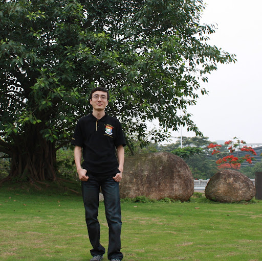
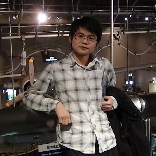
Liang Zhang
age ~40
from Issaquah, WA
- Also known as:
-
- Lian G Zhang
- Leec Zhang Liang
- Phone and address:
- 22902 SE 136Th Ct, Bellevue, WA 98027
Liang Zhang Phones & Addresses
- 22902 SE 136Th Ct, Issaquah, WA 98027
- Vancouver, WA
- Seattle, WA
- 38392 Midnight Sky Pl, Hamilton, VA 20158 • (571)3150458
- Leesburg, VA
- 38392 Midnight Sky Pl, Hamilton, VA 20158
Work
-
Company:Cell and molecular biology labJan 2013
-
Position:Lab instructor
Education
-
School / High School:Michigan State University- East Lansing, MIAug 2007
-
Specialities:Ph.D. in Cell and Molecular Biology
Ranks
-
Licence:New York - Due to reregister within 30 days of birthday
-
Date:2011
Specialities
Tax
Isbn (Books And Publications)

Proceedings of the Twentieth International Cryogenic Engineering Conference (ICEC 20): Beijing, China, 11-14 May 2004
view sourceAuthor
Liang Zhang
ISBN #
0080445594

La Naissance Du Concept De Patrimoine En Chine: XIX-XXe Siecles
view sourceAuthor
Liang Zhang
ISBN #
2862220477
Medicine Doctors

Liang Zhang
view sourceSpecialties:
Anesthesiology
Work:
Covenant Home Center Operating Room
3421 W 9 St FL 2, Waterloo, IA 50702
(319)2728000 (phone), (319)2727313 (fax)
3421 W 9 St FL 2, Waterloo, IA 50702
(319)2728000 (phone), (319)2727313 (fax)
Languages:
English
Description:
Dr. Zhang works in Waterloo, IA and specializes in Anesthesiology. Dr. Zhang is affiliated with Covenant Medical Center.

Liang Zhang
view source
Liang Zhang
view sourceName / Title
Company / Classification
Phones & Addresses
Chairman of the Board
Synutra International, Inc
Mfg Canned Specialties · Mfg Dairy Based Nutritional Products
Mfg Canned Specialties · Mfg Dairy Based Nutritional Products
2275 Res Blvd, Rockville, MD 20850
2275 Research Blvd SUITE 500, Rockville, MD 20850
(301)8403888
2275 Research Blvd SUITE 500, Rockville, MD 20850
(301)8403888
JACKIE DRAGON LLC
JACKIE MISO, LLC
Us Patents
-
Device, Method And System For Optical Communication With A Waveguide Structure And An Integrated Optical Coupler Of A Photonic Integrated Circuit Chip
view source -
US Patent:20220413210, Dec 29, 2022
-
Filed:Jun 25, 2021
-
Appl. No.:17/359178
-
Inventors:- Santa Clara CA, US
Pooya Tadayon - Portland OR, US
Zhichao Zhang - Chandler AZ, US
Liang Zhang - Chandler AZ, US -
Assignee:Intel Corporation - Santa Clara CA
-
International Classification:G02B 6/122
G02B 6/30 -
Abstract:Techniques and mechanisms for optically coupling a photonic integrated circuit (PIC) chip to an optical fiber via a planar optical waveguide structure. In an embodiment, a PIC chip comprises integrated circuitry, photonic waveguides, and integrated edge-oriented couplers (IECs) which are coupled to the integrated circuitry via the photonic waveguides. The PIC chip forms respective divergent lens surfaces of the IECs, which are each at a respective terminus of a corresponding one of the photonic waveguides. A planar optical waveguide structure, which is adjacent to the IECs, comprises a core which is optically coupled between the PIC chip and an array of optical fibers. In another embodiment, an edge of the PIC forms a stepped structure, wherein an upper portion of the stepped structure comprises the plurality of coplanar IECs, and a lower portion of the stepped structure extends past the plurality of coplanar IECs.
-
Device, Method And System For Optical Communication With A Photonic Integrated Circuit Chip And A Transverse Oriented Lens Structure
view source -
US Patent:20220413214, Dec 29, 2022
-
Filed:Jun 25, 2021
-
Appl. No.:17/359183
-
Inventors:- Santa Clara CA, US
Pooya Tadayon - Portland OR, US
Zhichao Zhang - Chandler AZ, US
Liang Zhang - Chandler AZ, US
Srikant Nekkanty - Chandler AZ, US -
Assignee:Intel Corporation - Santa Clara CA
-
International Classification:G02B 6/12
G02B 3/06 -
Abstract:Techniques and mechanisms for facilitating horizontal communication with a photonic integrated circuit (PIC) chip, and a lens structure which is optically coupled thereto. In an embodiment, a PIC chip comprises integrated circuitry, photonic waveguides, and integrated edge-oriented couplers (IECs) which are coupled to the integrated circuitry via the photonic waveguides. The PIC chip forms respective first divergent lens surfaces of the IECs, which are each at a respective terminus of a corresponding one of the photonic waveguides. A lens structure, which is adjacent to the IECs, comprises a second divergent lens surface having an orientation which is substantially orthogonal to the respective orientations of the first divergent lens surfaces. In another embodiment, an edge of the PIC chip forms one or more recess structures, and the lens structure comprises one or more tenon portions which each extends into a respective recess structure of the one or more recess structures.
-
Proximity-Based Unlocking Of Communal Computing Devices
view source -
US Patent:20220321561, Oct 6, 2022
-
Filed:Jun 22, 2022
-
Appl. No.:17/808172
-
Inventors:- Redmond WA, US
Dipesh BHATTARAI - Redmond WA, US
Peter Gregory DAVIS - Redmond WA, US
Jeffrey JOHNSON - Bellevue WA, US
Liang ZHANG - Redmond WA, US
Kiran KUMAR - Redmond WA, US -
Assignee:Microsoft Technology Licensing, LLC - Redmond WA
-
International Classification:H04L 9/40
H04W 12/63 -
Abstract:A communal computing device, such as an interactive digital whiteboard, can become unlocked if a user is near the device. The communal computing device may use a sensor such as a camera to capture images of a person and obtain an identifier from a personal device such as a smartphone. A cloud-based provider that is trusted by both the communal computing device and the personal device may associate both the image and the identifier of the personal device with the same user identity. Obtaining the user identity from multiple, different sources provides a secure technique for the communal computing device to recognize a user without the user directly interacting with the communal computing device. If the sensor no longer detects the user or the personal device is no longer detected, then the communal computing device may log off the user. The personal device may be used to confirm log off.
-
Three Dimensional Volume Flow Quantification And Measurement
view source -
US Patent:20220183655, Jun 16, 2022
-
Filed:Mar 19, 2020
-
Appl. No.:17/439830
-
Inventors:- EINDHOVEN, NL
JAMES ROBERTSON JAGO - SEATTLE WA, US
SIBO LI - WALTHAM MA, US
SHIYING WANG - MELROSE MA, US
JUN SOEB SHIN - WINCHESTER MA, US
GERARD JOSEPH HARRISON - SNOHOMISH WA, US
THANASIS LOUPAS - KIRKLAND WA, US
LIANG ZHANG - ISSAQUAH WA, US -
International Classification:A61B 8/06
A61B 8/08
A61B 8/02
A61B 8/00 -
Abstract:An ultrasonic diagnostic imaging system acquires volume image flow data sets of subvolumes of a blood vessel over at least a cardiac cycle. Image data of the subvolumes is then aligned both spatially and temporally to produce 3D images of the volume flow of the blood vessel over a heart cycle. A volume flow profile curve is produced from the acquired volume image flow data sets. The subvolumes are scanned starting with the center of the blood vessel and proceeding outward therefrom. The blood vessel center may be designated manually by a user or automatically by the ultrasound system by Doppler or other methods. Each subvolume is scanned over a heart cycle, with the systolic phase in the temporal center of the acquisition interval. The subvolumes are scanned in synchronism with the heart cycle and the estimation of a heart cycle is updated during each subvolume data acquisition interval.
-
Systems And Methods For Automatic Detection And Visualization Of Turbulent Blood Flow Using Vector Flow Data
view source -
US Patent:20230085700, Mar 23, 2023
-
Filed:Nov 22, 2022
-
Appl. No.:17/992423
-
Inventors:- Eindhoven, NL
Shiying Wang - Melrose MA, US
Sheng-Wen Huang - Ossining NY, US
Francois Guy Gerard Marie Vignon - Andover MA, US
Keith William Johnson - Lynnwood WA, US
Liang Zhang - Issaquah WA, US
David Hope Simpson - Bothell WA, US -
International Classification:A61B 8/06
A61B 8/00
A61B 8/08 -
Abstract:A system for visualization and quantification of ultrasound imaging data according to embodiments of the present disclosure may include a display unit, and a processor communicatively coupled to the display unit and to an ultrasound imaging apparatus for generating an image from ultrasound data representative of a bodily structure and fluid flowing within the bodily structure. The processor may be configured to estimate axial and lateral velocity components of the fluid flowing within the bodily structure, determine a plurality of flow directions within the image based on the axial and lateral velocity components, differentially encode the flow directions based on flow direction angle to generate a flow direction map, and cause the display unit to concurrently display the image including the bodily structure overlaid with the flow direction map.
-
Systems And Methods For Vascular Rendering
view source -
US Patent:20230073704, Mar 9, 2023
-
Filed:Mar 5, 2021
-
Appl. No.:17/908341
-
Inventors:- EINDHOVEN, NL
Paul Sheeran - Woodinville WA, US
Thanasis Loupas - Kirkland WA, US
Liang Zhang - Issaquah WA, US -
International Classification:G06T 11/00
G06T 5/20
G06T 5/00
G06T 7/90
G06T 5/50 -
Abstract:In some examples, color Doppler data may be separated into luminance data and chrominance data. The luminance data may be modified without modifying the chrominance data. In some examples, the luminance data may be adjusted based, at least in part, on power Doppler data. The adjusted luminance data may be recombined with the chrominance data to provide augmented color Doppler data. In some examples, the power Doppler data may be enhanced by filtering, for example, by applying a Frangi vesselness filter, prior to being used to adjust the luminance data of the color Doppler data.
-
Adaptive Ultrasound Flow Imaging
view source -
US Patent:20210373154, Dec 2, 2021
-
Filed:Oct 14, 2019
-
Appl. No.:17/287623
-
Inventors:- EINDHOVEN, NL
SHENG-WEN HUANG - OSSINING NY, US
HUA XIE - CAMBRIDGE MA, US
KEITH WILLIAM JOHNSON - LYNWOOD WA, US
LIANG ZHANG - ISSAQUAH WA, US
THANASIS LOUPAS - KIRKLAND WA, US
TRUONG HUY NGUYEN - REDMOND WA, US -
International Classification:G01S 15/89
A61B 8/06
A61B 8/08 -
Abstract:The present disclosure describes ultrasound systems configured to enhance flow imaging and analysis by adaptively adjusting one or more imaging parameters in response to acquired flow measurements. Example systems can include an ultrasound transducer and one or more processors. Using the system components, mean flow velocity magnitude and acceleration can be determined within a target region during an acquisition phase, which may include a cardiac cycle. One or more adjusted flow imaging parameters, such as adjusted ensemble length, temporal smoothing filter length and/or step size, can be determined based on the acquired flow measurements to increase the signal quality of newly acquired ultrasound echo signals. The adjusted flow imaging parameters can then be applied by the ultrasound transducer during a second acquisition phase.
-
Systems And Methods For Automatic Detection And Visualization Of Turbulent Blood Flow Using Vector Flow Data
view source -
US Patent:20210145399, May 20, 2021
-
Filed:May 25, 2018
-
Appl. No.:16/616753
-
Inventors:- EINDHOVEN, NL
SHIYING WANG - MELROSE MA, US
SHENG-WEN HUANG - OSSINING NY, US
FRANCOIS GUY GERARD MARIE VIGNON - ANDOVER MA, US
KEITH WILLIAM JOHNSON - LYNWOOD WA, US
LIANG ZHANG - ISSAQUAH WA, US
DAVID HOPE SIMPSON - BOTHELL WA, US -
Assignee:KONINKLIJKE PHILIPS N.V. - EINDHOVEN
-
International Classification:A61B 8/06
A61B 8/00
A61B 8/08 -
Abstract:A system for visualization and quantification of ultrasound imaging data according to embodiments of the present disclosure may include a display unit, and a processor communicatively coupled to the display unit and to an ultrasound imaging apparatus for generating an image from ultrasound data representative of a bodily structure and fluid flowing within the bodily structure. The processor may be configured to estimate axial and lateral velocity components of the fluid flowing within the bodily structure, determine a plurality of flow directions within the image based on the axial and lateral velocity components, differentially encode the flow directions based on flow direction angle to generate a flow direction map, and cause the display unit to concurrently display the image including the bodily structure overlaid with the flow direction map.
Lawyers & Attorneys

Liang Zhang - Lawyer
view sourceAddress:
Shulun & Partners (Shanghai) Law Firm
Licenses:
New York - Due to reregister within 30 days of birthday 2011
Education:
Chicago-Kent College of Law, Iit

Liang Zhang - Lawyer
view sourceOffice:
Davis Polk & Wardwell LLP
Specialties:
Tax
ISLN:
1000248654
Admitted:
2017
University:
Lafayette College, B.S., 2013
Law School:
Harvard Law School, J.D., 2016
Resumes

Liang Zhang Redmond, WA
view sourceWork:
Cell and Molecular Biology Lab
Jan 2013 to May 2013
Lab instructor Michigan State University
Aug 2007 to May 2013
Research assistant Eukaryotic Cell Biology
Jan 2009 to May 2009
Teaching assistant Sichuan University
Sep 2003 to Jul 2007
Undergraduate researcher University of Washington
Sep 2005 to Jul 2006
Undergraduate researcher
Education:
Michigan State University
East Lansing, MI
Aug 2007 to May 2013
Ph.D. in Cell and Molecular Biology Sichuan University
Chengdu, CN
Sep 2003 to Jul 2007
Bachelor of Science in Biology University of Washington
Seattle, WA
Sep 2005 to Jul 2006
East Lansing, MI
Aug 2007 to May 2013
Ph.D. in Cell and Molecular Biology Sichuan University
Chengdu, CN
Sep 2003 to Jul 2007
Bachelor of Science in Biology University of Washington
Seattle, WA
Sep 2005 to Jul 2006
Myspace
Googleplus

Liang Zhang
Education:
Universiteit van Tilburg, Peking University, Harbin Institute of Technology
Tagline:
I have an attitude and I know how to use it.

Liang Zhang
Education:
Harbin Institute of Technology

Liang Zhang
Work:
Spun Tech Industry - Summer Intern
Education:
North Carolina State University

Liang Zhang
Education:
Yale University

Liang Zhang
Education:
Yale University

Liang Zhang
Education:
Huazhong University of Science and Technology

Liang Zhang

Liang Zhang
Flickr
Plaxo

Zhang Liang
view sourceNottingham, UKPast: Nokia Siemens Networks, Siemens Communications, Huawei Technologies, UTStarcom, ...

zhang liang
view sourcehangzhou一个感性的完美主义者
Classmates

Liang Zhang
view sourceSchools:
Albert Enstine High School Wheaton MD 1993-1997
Community:
Frances Hunt, Sonia Carpenter, Stuart Harris, Barry Mitzner

Liang Zhang
view sourceSchools:
Shanghai American School Shanghai China 1998-2000
Community:
David Giedt, Glenda Case, Raazia Bokhari, Eugene Wu

Liang Zhang
view sourceSchools:
Zhejiang University Hangzhou China 1993-1997
Community:
Lisa Chen, Feng Feng, Dongwei Wang, Wu Hai

Liang Zhang (Hongchen)
view sourceSchools:
Beijing Bayi High School Beijing China 1992-1996

Liang Zhang
view sourceSchools:
Cleveland Hill Elementary School Cheektowaga NY 1993-1996
Youtube

Zhang Dg Liang
view source
Dg Liang Zhang
view source
Liang Ivy Zhang
view source
Zhang Liang
view source
Liang Tom Zhang
view source
Lim Liang Zhang
view source
Wen Liang Zhang
view source
Liang Zhang
view sourceGet Report for Liang Zhang from Issaquah, WA, age ~40


