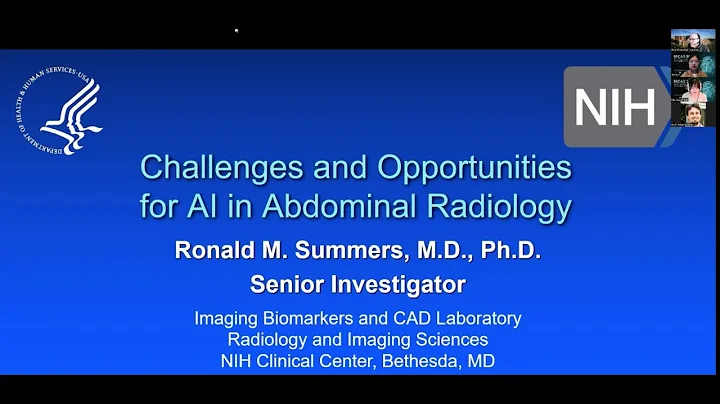
Ronald A Summers
age ~55
from Alexandria, VA
- Also known as:
-
- Ron Summers
- Phone and address:
-
3610 Lakota Rd, Alexandria, VA 22303
(703)9609873
Ronald Summers Phones & Addresses
- 3610 Lakota Rd, Alexandria, VA 22303 • (703)9609873
- 8101 Clifforest Dr, Springfield, VA 22153
- 6911 Spelman Dr, Springfield, VA 22153 • (703)6445054
- Woodbridge, VA
Education
-
School / High School:Perelman School of Medicine University of Pennsylvania1988
Languages
English
Awards
Healthgrades Honor Roll
Ranks
-
Certificate:Diagnostic Radiology, 1993
Specialities
Diagnostic Radiology
Medicine Doctors

Dr. Ronald M Summers, Bethesda MD - MD (Doctor of Medicine)
view sourceSpecialties:
Diagnostic Radiology
Address:
9000 Rockville Pike Suite 10, Bethesda, MD 20892
(301)4025486 (Phone)
1500 E Medical Center Dr, Ann Arbor, MI 48109
(301)4025486 (Phone)
1500 E Medical Center Dr, Ann Arbor, MI 48109
Certifications:
Diagnostic Radiology, 1993
Awards:
Healthgrades Honor Roll
Languages:
English
Education:
Medical School
Perelman School of Medicine University of Pennsylvania
Graduated: 1988
Medical School
Presby Med Ctr
Graduated: 1988
Medical School
University Of Michigan Hospitals
Graduated: 1988
Medical School
Duke University Hospital
Graduated: 1988
Perelman School of Medicine University of Pennsylvania
Graduated: 1988
Medical School
Presby Med Ctr
Graduated: 1988
Medical School
University Of Michigan Hospitals
Graduated: 1988
Medical School
Duke University Hospital
Graduated: 1988

Ronald A. Summers
view sourceSpecialties:
Orthopaedic Surgery, Orthopedic Sports Medicine
Work:
Wake Orthopaedics LLC
3009 New Bern Ave, Raleigh, NC 27610
(919)2325020 (phone), (919)2325021 (fax)
Wake Orthopaedics LLC
212 Ashville Ave STE 30, Cary, NC 27518
(919)2350616 (phone), (919)2350610 (fax)
Wake Orthopaedics LLC
8001 T W Alexander Dr STE 224, Raleigh, NC 27617
(919)7147152 (phone), (919)2325021 (fax)
3009 New Bern Ave, Raleigh, NC 27610
(919)2325020 (phone), (919)2325021 (fax)
Wake Orthopaedics LLC
212 Ashville Ave STE 30, Cary, NC 27518
(919)2350616 (phone), (919)2350610 (fax)
Wake Orthopaedics LLC
8001 T W Alexander Dr STE 224, Raleigh, NC 27617
(919)7147152 (phone), (919)2325021 (fax)
Education:
Medical School
University of Kansas School of Medicine
Graduated: 1992
University of Kansas School of Medicine
Graduated: 1992
Procedures:
Hip/Femur Fractures and Dislocations
Arthrocentesis
Carpal Tunnel Decompression
Hallux Valgus Repair
Hip Replacement
Joint Arthroscopy
Knee Arthroscopy
Knee Replacement
Lower Arm/Elbow/Wrist Fractures and Dislocations
Lower Leg/Ankle Fractures and Dislocations
Occupational Therapy Evaluation
Shoulder Arthroscopy
Shoulder Surgery
Arthrocentesis
Carpal Tunnel Decompression
Hallux Valgus Repair
Hip Replacement
Joint Arthroscopy
Knee Arthroscopy
Knee Replacement
Lower Arm/Elbow/Wrist Fractures and Dislocations
Lower Leg/Ankle Fractures and Dislocations
Occupational Therapy Evaluation
Shoulder Arthroscopy
Shoulder Surgery
Conditions:
Internal Derangement of Knee
Intervertebral Disc Degeneration
Rotator Cuff Syndrome and Allied Disorders
Fractures, Dislocations, Derangement, and Sprains
Internal Derangement of Knee Cartilage
Intervertebral Disc Degeneration
Rotator Cuff Syndrome and Allied Disorders
Fractures, Dislocations, Derangement, and Sprains
Internal Derangement of Knee Cartilage
Languages:
English
Spanish
Spanish
Description:
Dr. Summers graduated from the University of Kansas School of Medicine in 1992. He works in Raleigh, NC and 2 other locations and specializes in Orthopaedic Surgery and Orthopedic Sports Medicine. Dr. Summers is affiliated with Wakemed Cary Hospital and Wakemed Raleigh Campus.
Us Patents
-
Method For Segmenting Medical Images And Detecting Surface Anomalies In Anatomical Structures
view source -
US Patent:6345112, Feb 5, 2002
-
Filed:Jan 19, 2001
-
Appl. No.:09/765826
-
Inventors:Ronald M. Summers - Potomac MD
Scott Selbie - Rockville MD
James D. Malley - Rockville MD
Lynne M. Pusanik - Columbia MD -
Assignee:The United States of America as represented by the Department of Health and Human Services - Washington DC
-
International Classification:G06K 900
-
US Classification:382128
-
Abstract:A region growing method segments three-dimensional image data of an anatomical structure using a tortuous path length limit to constrain voxel growth. The path length limit constrains the number of successive generations of voxel growth from a seed point to prevent leakage of voxels outside the boundary of the anatomical structure. Once segmented, a process for detecting surface anomalies performs a curvature analysis on a computer model of the surface of the structure. This process detects surface anomalies automatically by traversing the vertices in the surface model, computing partial derivatives of the surface at the vertices, and computing curvature characteristics from the partial derivatives. To identify possible anomalies, the process compares the curvature characteristics with predetermined curvature characteristics of anomalies and classifies the vertices. The process further refines potential anomalies by segmenting neighboring vertices that are classified as being part of an anomaly using curvature characteristics.
-
Method For Segmenting Medical Images And Detecting Surface Anomalies In Anatomical Structures
view source -
US Patent:6556696, Apr 29, 2003
-
Filed:Feb 5, 2002
-
Appl. No.:10/072667
-
Inventors:Ronald M. Summers - Potomac MD
Scott Selbie - Rockville MD
James D. Malley - Rockville MD
Lynne M. Pusanik - Columbia MD -
Assignee:The United States of America as represented by the Department of Health and Human Services - Washington DC
-
International Classification:G06K 900
-
US Classification:382128
-
Abstract:A region growing method segments three-dimensional image data of an anatomical structure using a tortuous path length limit to constrain voxel growth. The path length limit constrains the number of successive generations of voxel growth from a seed point to prevent leakage of voxels outside the boundary of the anatomical structure. Once segmented, a process for detecting surface anomalies performs a curvature analysis on a computer model of the surface of the structure. This process detects surface anomalies automatically by traversing the vertices in the surface model, computing partial derivatives of the surface at the vertices, and computing curvature characteristics from the partial derivatives. To identify possible anomalies, the process compares the curvature characteristics with predetermined curvature characteristics of anomalies and classifies the vertices. The process further refines potential anomalies by segmenting neighboring vertices that are classified as being part of an anomaly using curvature characteristics.
-
Computer-Aided Classification Of Anomalies In Anatomical Structures
view source -
US Patent:7260250, Aug 21, 2007
-
Filed:Sep 26, 2003
-
Appl. No.:10/671749
-
Inventors:Ronald M. Summers - Potomac MD, US
Marek Franaszek - Gaithersburg MD, US
Gheorghe Iordanescu - Rockville MD, US -
Assignee:The United States of America as represented by the Secretary of the Department of Health and Human Services - Washington DC
-
International Classification:G06K 9/00
-
US Classification:382128, 382286
-
Abstract:Candidate anomalies in an anatomical structure are processed for classification. For example, false positives can be reduced by techniques related to the anomaly's neck, wall thickness associated with the anomaly, template matching performed for the anomaly, or some combination thereof. The various techniques can be combined for use in a classifier, which can determine whether the anomaly is of interest. For example, a computed tomography scan of a colon can be analyzed to determine whether a candidate anomaly is a polyp. The technologies can be applied to a variety of other scenarios involving other anatomical structures.
-
Automated Identification Of Ileocecal Valve
view source -
US Patent:7440601, Oct 21, 2008
-
Filed:Oct 8, 2004
-
Appl. No.:10/961681
-
Inventors:Ronald Summers - Potomac MD, US
Jianhua Yao - Columbia MD, US
C. Daniel Johnson - Rochester MD, US -
Assignee:The United States of America as represented by the Department of Health and Human Services - Washington DC
Mayo Foundation for Medical Education and Research - Rochester MN -
International Classification:G06K 9/00
-
US Classification:382128, 382131, 382132
-
Abstract:Portions of a virtual colon can be analyzed to identify a normal structure, such as an ileocecal valve. Paradigmatic characteristics of ileocecal valves can be used to identify a digital representation as an ileocecal valve. Upon determination that a digital representation has the characteristics of an ileocecal valve, action can be taken. For example, the digital representation can be removed from a list of polyp candidates.
-
Determination Of Feature Boundaries In A Digital Representation Of An Anatomical Structure
view source -
US Patent:7454045, Nov 18, 2008
-
Filed:Feb 13, 2004
-
Appl. No.:10/779210
-
Inventors:Jianhua Yao - Laurel MD, US
Ronald M. Summers - Potomac MD, US -
Assignee:The United States of America as represented by the Department of Health and Human Services - Washington DC
-
International Classification:G06K 9/00
-
US Classification:382128, 382130, 382131, 382132, 382154, 382224
-
Abstract:A virtual anatomical structure can be analyzed to determine enclosing three-dimensional boundaries of features therein. Various techniques can be used to determine tissue types in the virtual anatomical structure. For example, tissue types can be determined via an iso-boundary between lumen and air in the virtual anatomical structure and a fuzzy clustering approach. Based on the tissue type determination, a deformable model approach can be used to determine an enclosing three-dimensional boundary of a feature in the virtual anatomical structure. The enclosing three-dimensional boundary can be used to determine characteristics of the feature and classify it as of interest or not of interest.
-
Automated Centerline Detection Algorithm For Colon-Like 3D Surfaces
view source -
US Patent:7570802, Aug 4, 2009
-
Filed:Dec 18, 2002
-
Appl. No.:10/500342
-
Inventors:Gheorghe Iordanescu - Rockville MD, US
Ronald M. Summers - Potomac MD, US
Juan Raul Cebral - Vienna VA, US -
Assignee:The United States of America as represented by the Secretary of the Department of Health and Human Services - Washington DC
-
International Classification:G06K 9/00
-
US Classification:382154, 382128, 382130, 382131, 382133, 600407
-
Abstract:A three dimensional image of the colon like surface is processed to determine at least its ring structure. The image is composed of vertex points, each vertex point having a discrete point identifier and three dimensional position information. The three dimensional position information is averaged in a shrinking procedure to contract the three dimensional image proximate to a major axis of the colon-like surface. Evenly spaced points are taken through the shrunken colon like surface and connected to form a curve. Planes are generated at intervals normal to the curve to split the shrunken colon like surface into image segments. By mapping these image segments back to the original image through their discrete point identifiers, an accurate ring profile of the colon like surface can be generated.
-
Teniae Coli Guided Navigation And Registration For Virtual Colonoscopy
view source -
US Patent:7570986, Aug 4, 2009
-
Filed:May 17, 2006
-
Appl. No.:11/436889
-
Inventors:Hui-Yang Huang - Rockville MD, US
Dave A. Roy - Dallas TX, US
Ronald M. Summers - Potomac MD, US -
Assignee:The United States of America as represented by the Secretary of Health and Human Services - Washington DC
-
International Classification:A61B 5/05
-
US Classification:600425, 600407
-
Abstract:A computer-assisted method for detecting surface features in a virtual colonoscopy. The method includes providing a three-dimensional construction of a computed tomography colonography surface; creating a path along the teniae coli from the proximal ascending colon to the distal descending colon on the colonography surface; forming an indexed computed tomography colonography surface using the created path; and registering the supine and prone scans of the computed tomography colonography surface using the indexed computed tomography colonography surface. The method also includes navigating the internal surface of the computed tomography colonography using the indexed computed tomography colonography surface.
-
Computer-Aided Classification Of Anomalies In Anatomical Structures
view source -
US Patent:7646904, Jan 12, 2010
-
Filed:Jul 9, 2007
-
Appl. No.:11/775011
-
Inventors:Ronald M. Summers - Potomac MD, US
Marek Franaszek - Gaithersburg MD, US
Gheorghe Iordanescu - Rockville MD, US -
Assignee:The United States of America as represented by the Department of Health and Human Services - Washington DC
-
International Classification:G06K 9/00
-
US Classification:382128
-
Abstract:Candidate anomalies in an anatomical structure are processed for classification. For example, false positives can be reduced by techniques related to the anomaly's neck, wall thickness associated with the anomaly, template matching performed for the anomaly, or some combination thereof. The various techniques can be combined for use in a classifier, which can determine whether the anomaly is of interest. For example, a computed tomography scan of a colon can be analyzed to determine whether a candidate anomaly is a polyp. The technologies can be applied to a variety of other scenarios involving other anatomical structures.
Name / Title
Company / Classification
Phones & Addresses
RON SUMMERS ROOFING, LLC
Vice President
COLUMBIA RIVER SAND & GRAVEL, INC
Isbn (Books And Publications)

ITAB 2003: Conference Proceedings, 4th International IEEE EMBS Special Topic Conference on Information Technology Applications in Biomedicine, 24-26 April 2003, Birmingham, United Kingdom New Solutio
view sourceAuthor
Ronald Summers
ISBN #
0780376676
License Records
Ronald Alan Summers Md
License #:
2873 - Expired
Category:
Medicine
Issued Date:
Jul 1, 1992
Effective Date:
Jul 2, 1993
Type:
Temporary Educational Permit
Resumes

Ronald Summers
view source
Ronald Summers
view source
Ronald Summers
view sourceSkills:
Management
Microsoft Excel
Microsoft Office
Research
Microsoft Excel
Microsoft Office
Research

Ronald Summers
view source
Ronald Summers
view source
Ronald Summers
view sourceLocation:
United States

Ronald Summers
view sourceLocation:
United States

Ronald Summers
view sourceLocation:
United States

Ronald Summers
view source
Ronald Summers
view source
Ronald Summers
view source
Ronald Summers
view source
Ronald Summers
view source
Ronald C Summers
view source
Ronald Summers
view source
Ronald D Summers
view sourceClassmates

Ronald Summers
view sourceSchools:
Florida Central Academy Sorrento FL 1976-1977
Community:
Laurie Friese, Laura Anderson, Pamela Waugh, Henry Hamilton, Gail Campbell

Ronald Summers
view sourceSchools:
Gonzaga High School St. John's Peru 1965-1969
Community:
Matt Gauci, Cathy Gillies, Darren Walker

Ronald Summers
view sourceSchools:
Florida Central Academy Sorrento FL 1973-1977
Community:
Laurie Friese, Laura Anderson, Pamela Waugh, Henry Hamilton, Gail Campbell

Ronald Summers
view sourceSchools:
George Rogers Clark School 1 Indianapolis IN 1953-1957
Community:
Ted Levee, Mark Leavell, Darlene Barnett

Ronald Summers
view sourceSchools:
Thomastown School Akron OH 1953-1957
Community:
Pamela Sellers, Barbara Bellomy

Ronald Summers
view sourceSchools:
Fultondale High School Birmingham AL 1977-1981
Community:
Donna Thomas, Charlene Martin, Hollie Schmidt, Leon Reese

Ronald Summers | Branchvi...
view source
Ronald Summers, Okmulgee ...
view sourceFlickr
Googleplus

Ronald Summers

Ronald Summers
Youtube
Myspace

Ronald Summers
view sourceGender:
Male
Get Report for Ronald A Summers from Alexandria, VA, age ~55


